Difference Between Primary and Secondary Hemostasis
Key Difference – Primary vs Secondary Hemostasis
When there is an injury in the body, the blood is changed from a fluid state to solid state in order to prevent bleeding. This occurs via a natural process called hemostasis. Hemostasis can be defined as a physiological process which stops excessive bleeding after an injury in a blood vessel. It is a complex and highly regulated process to localize blood clotting only at the injury site. Hemostasis involves several factors such as vascular factors, platelet factors and coagulating proteins. The final outcome of hemostasis is the coagulation of blood at the wound site. Hemostasis occurs via two connected phases named primary hemostasis and secondary hemostasis. Hemostasis initiates with primary hemostasis. During primary hemostasis, platelets in the blood aggregate at the injury site and form a platelet plug to block the hole. Primary hemostasis is followed by secondary hemostasis. During secondary hemostasis, platelet plug is further reinforced by a fibrin mesh produced through proteolytic coagulation cascade. Therefore, the key difference between primary and secondary hemostasis is that primary hemostasis makes a weak platelet plug at the injury site while secondary hemostasis makes it strong by generating a fibrin mesh on it.
CONTENTS
1. Overview and Key Difference
2. What is Primary Hemostasis
3. What is Secondary Hemostasis
4. Side by Side Comparison – Primary vs Secondary Hemostasis in Tabular Form
5. Summary
What is Primary Hemostasis?
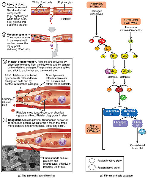
The endothelium of the blood vessels maintains an anticoagulating surface inside the blood vessels to maintain blood fluidity. However, when there is an injury in a blood vessel, several components in the subendothelial matrix activate and initiate the formation of a blood clot around the injury. This process is known as hemostasis. Hemostasis has two phases. During the first phase of hemostasis, platelets in the blood aggregate and form a platelet plug to block the open hole in the blood vessel. This phase is known as primary hemostasis. Platelets are activated through a series of biological processes and, as a result, they adhere at the site of injury and aggregate to each other to form a plug.
Primary hemostasis starts immediately after the blood vessel disruption. The blood vessel near the injury site contracts temporally to narrow it and reduce the blood flow. This is the first step of primary hemostasis and it is known as vasoconstriction. It reduces the amount of blood loss and enhances the platelet adherence and activation at the wound site. When the platelets are activated, they attract other platelets to form a plug to block the opening. Vasoconstriction can be achieved by two ways: through nerve system or through the molecules called endothelin secreted by endothelial cells.

Figure 01: Hemostasis Process
Platelet adhesion is supported by different types of molecules such as glycoproteins located on platelets, collagens, and von Willebrand factor (vWf). Glycoproteins of the platelets adhere to vWf, which is a sticky molecule. Then these platelets collect at the injury site and activate upon the contraction with collagen. Collagen activated platelets form pseudopods which distribute to cover the injury surface. Then fibrinogen binds with receptors at the collagen-activated platelets. Fibrinogen provides more sites for platelet to bind with each other. Hence, other platelets are also aggregated at the injury surface and make a soft platelet plug over the injury hole.
What is Secondary Hemostasis?
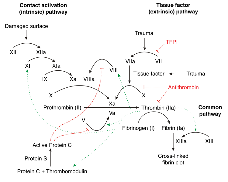
Secondary hemostasis is the second phase of hemostasis. During secondary hemostasis, the soft platelet plug formed during the primary hemostasis is made stronger by the formation of a fibrin mesh on it. Fibrin is an insoluble plasma protein which serves as the underlying fabric polymer of a blood clot. Fibrin mesh strengthens and stabilizes the soft platelet plug formed at the injury site. Fibrin formation occurs via coagulation factors through coagulation cascade.

Figure 02: Formation of fibrin clot by secondary hemostasis
Different kinds of coagulation factors are synthesized by the liver and released into the blood. Initially, they are inactive and later become activated by subendothelial collagens or by thromboplastin. Subendothelial collagen and thromboplastin are released due to an injury occurred in the endothelium of the blood vessel. When they are released into the blood, they activate coagulation factors in the blood. These coagulation factors are activated one after the other, and finally, convert fibrinogen into fibrin. Then the fibrin links on the top of the platelet plug and makes a mesh by making platelet plug stronger. Fibrin, together with platelet plug, forms a blood clot at the end of the hemostasis process.
What is the Difference Between Primary and Secondary Hemostasis?
Primary vs Secondary Hemostasis | |
| Primary hemostasis is the first phase of hemostasis. | Secondary hemostasis is the second phase of hemostasis. |
| Process | |
| Vascular contraction, platelet adhesion and formation of a platelet plug occur during primary hemostasis. | During secondary hemostasis, coagulation factors are activated and fibrinogen is converted into fibrin, forming a fibrin mesh. |
| Goal | |
| The goal of primary hemostasis is to form a platelet plug. | The goal of secondary hemostasis is to make the platelet plug stronger by linking fibrin together on the top of the platelet plug and make a mesh. |
| Components Involved | |
| Primary hemostasis involves platelets, glycoprotein receptors of the platelets, collagen, vWf, and fibrinogen. | Secondary hemostasis involves subendothelial collagen, thromboplastin, coagulation factors, fibrinogen, and fibrin. |
| Duration | |
| Primary hemostasis occurs over a short period of time. | Secondary hemostasis takes comparatively longer time duration. |
Summary – Primary vs Secondary Hemostasis
Hemostasis is the physiological process which prevents bleeding at the site of an injury while maintaining a normal blood flow at the other places of the circulation. Blood loss is stopped by the formation of a hemostatic plug at the injury site. Hemostasis occurs via two phases named primary and secondary hemostasis. Primary hemostasis immediately starts after the injury and creates a platelet plug over the injury surface. This platelet plug is reinforced by the conversion of fibrinogen into fibrin by coagulation cascade during the secondary hemostasis. This is the main difference between primary and secondary hemostasis.
Download PDF Version of Primary vs Secondary Hemostasis
You can download PDF version of this article and use it for offline purposes as per citation notes. Please download PDF version here Difference Between Primary and Secondary Hemostasis.
Image Courtesy:
1. “1909 Blood Clotting” By OpenStax College – Anatomy & Physiology, Connexions Web site. Jun 19, 2013., (CC BY 3.0) via Commons Wikimedia
2. “Coagulation full” By Joe D – Own work (CC BY-SA 3.0) via Commons Wikimedia
References:
1. Gale, Andrew J. “Current Understanding of Hemostasis.” Toxicologic pathology. U.S. National Library of Medicine, 2011. Web. Available here. 28 June 2017.
2.”Primary hemostasis.” Khan Academy. N.p., n.d. Web. Available here. 28 June 2017.
3.”Secondary hemostasis.” Khan Academy. N.p., n.d. Web. Available here. 28 June 2017.
ncG1vNJzZmivp6x7pbXFn5yrnZ6YsqOx07CcnqZemLyue8OinZ%2Bdopq7pLGMm5ytr5Wau2680aKkmqqpYq6vsIyvqmarlZi8r7DAq7BmoJWivLTAwKygrGc%3D